
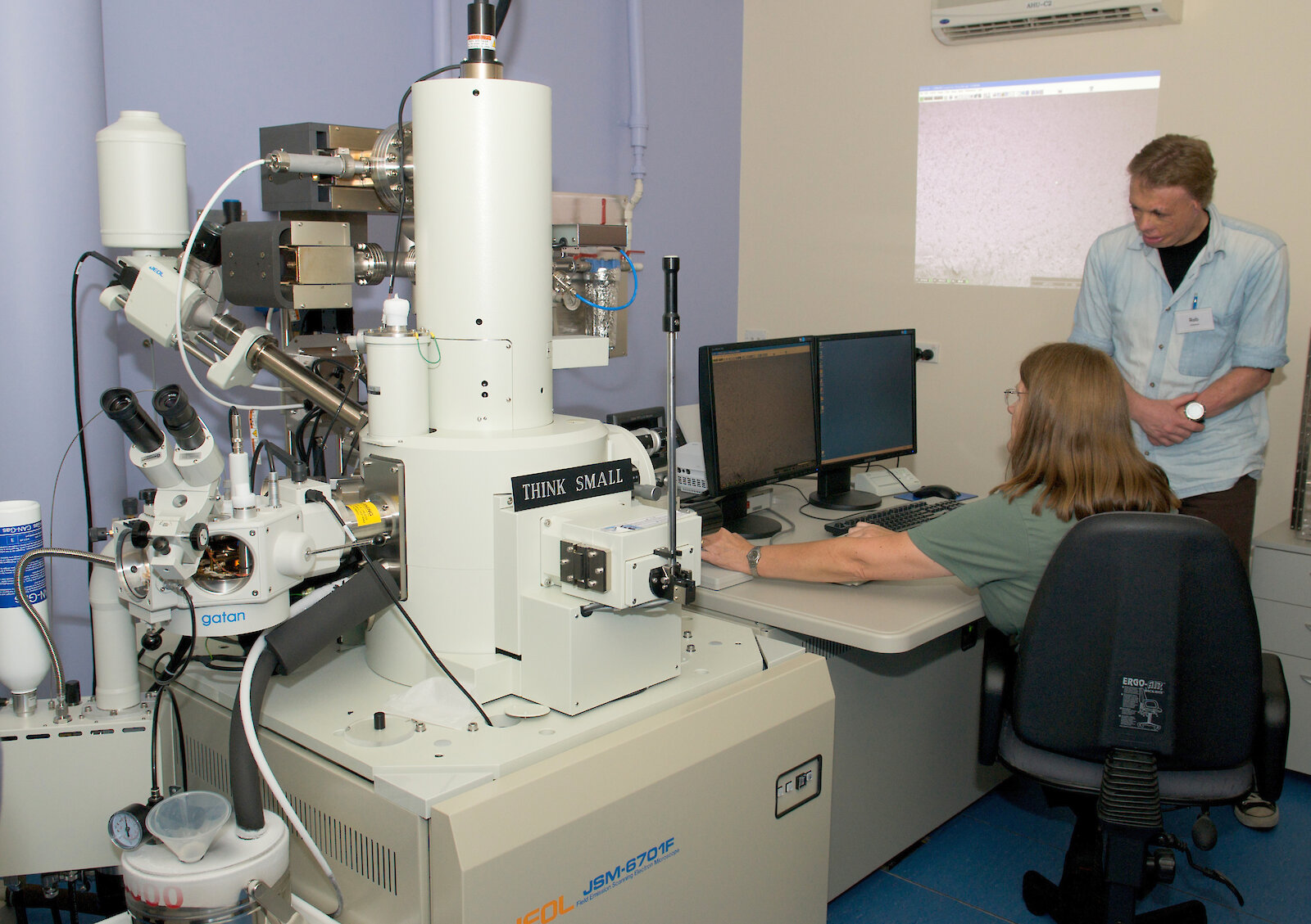
A new field emission Scanning Electron Microscope (SEM) using snap-freezing technology is helping Australian Antarctic Division scientists delve deeper into the biological mysteries of the microscopic world.
The $650,000 SEM produces detailed images of the surface structure of frozen specimens.
It magnifies to 650,000 times, allowing features two-million times smaller than the head of a pin to be observed.
Electron microscopist, Rick van den Enden, said the new SEM has a special ‘cryostage’ device that allows specimens to be imaged after they have been snap-frozen in super-cooled liquid nitrogen at minus 210 degrees Celsius.
“It used to take us almost a full day to prepare a specimen for imaging, but with the new “cryoSEM” specimens can be ready in just 20 minutes,” Mr van den Enden said.
“Importantly, the snap-frozen specimen is able to be observed in a more natural state with less damage and distortion, so we can get more accurate information.”
The Division’s microscopy work has traditionally focused on phytoplankton, krill, aging of seals by examining their teeth and fish by looking at their earbones.
“The new Scanning Electron Microscope and cryostage will allow us to extend our research to include delicate biological samples such as seaweeds and mosses which were not suitable for the previous equipment,” Mr van den Enden said.
A group of 20 international and Australian electron microscopists and other scientists are at the Antarctic Division this week, to learn more about the machine’s capabilities.
The new microscope is the only one of its type in Tasmania and will be available for use to the wider scientific community.